
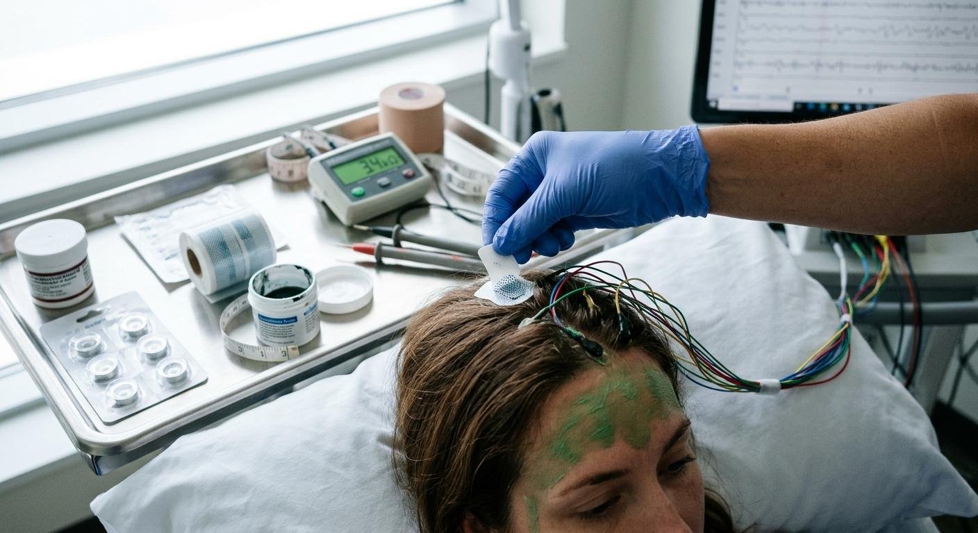

Understanding the Role of EEG Electrodes
EEG electrodes detect voltage differences produced by electrical activity in the brain. They are placed on the scalp according to a standardized electrode placement system, commonly the international 10-20 system or an expanded arrangement used for more detailed neurological monitoring.
These electrodes do not directly measure individual neurons. Instead, they detect electrical potentials generated by the synchronized activity of large groups of neurons, primarily through the scalp. The resulting signals are amplified, processed, and displayed as waveforms that clinicians interpret alongside the patient’s symptoms and clinical history.
Reliable measurements require a stable electrical connection between the electrode and the skin. Poor contact, dried conductive gel, movement, or inadequate preparation can introduce artifacts that make the recording harder to interpret.
Selecting appropriate eeg electrodes therefore involves more than choosing a particular material. The application environment, recording duration, patient population, and monitoring objectives all influence which option is most suitable.
Common Types of EEG Electrodes
Different electrode designs offer distinct advantages in terms of preparation time, signal stability, comfort, and suitability for prolonged monitoring.
Cup Electrodes
Cup electrodes are commonly used in clinical EEG examinations. They are typically made from conductive materials such as silver, silver chloride, gold, or other suitable metals, depending on the design and intended application.
Their shallow, cup-shaped structure holds conductive paste or gel against the scalp. Once secured, they can provide stable contact for routine EEG recordings and many diagnostic applications.
Advantages of cup electrodes include:
- Suitability for standard scalp EEG examinations.
- Reusability when the specific product is designed for it and appropriate cleaning and disinfection procedures are followed.
- Flexibility in placement according to standardized scalp landmarks.
- Compatibility with different conductive pastes and gels, subject to manufacturer instructions.
However, cup electrodes usually require careful scalp preparation and individual application. Technicians may need to separate hair, clean the skin, apply conductive material, and secure each electrode. This can make setup time-consuming when many channels are required.
Cup electrodes may be a good choice when precise placement and dependable contact are priorities. For repeated clinical use, proper inspection, reprocessing, and storage are essential to preserve electrode performance and patient safety.
Disposable EEG Electrodes
Disposable EEG electrodes are designed for single-use applications. They may incorporate conductive metal sensors, pre-applied conductive media, adhesive backing, or other features intended to simplify placement.
These electrodes are useful in environments where rapid setup, infection prevention, and efficient patient turnover are important. Depending on their design, they can reduce the time needed to prepare individual electrodes and eliminate the need to reprocess reusable components.
Potential benefits include:
- Consistent application when the electrodes are used according to instructions.
- Reduced cross-contamination risk when single-use products are handled and discarded properly.
- Convenient setup for routine monitoring and selected bedside applications.
- Less equipment reprocessing between patients.
Disposable designs are not automatically suitable for every EEG application. Some products are intended for short recordings, while others are developed for specific monitoring durations or environments. Clinicians should check the product specifications, approved application sites, connector compatibility, and recommended wear time before use.
It is also important to distinguish disposable EEG electrodes from adhesive ECG electrodes. Although both may use conductive sensors and adhesive materials, they are not necessarily interchangeable because their intended applications and performance requirements can differ.
Adhesive-Hydrogel EEG Electrodes
Adhesive-hydrogel electrodes combine an adhesive layer with a conductive hydrogel or similar conductive medium. The adhesive helps maintain contact with the scalp, while the conductive material supports electrical coupling between the skin and the sensor.
These electrodes can be helpful when monitoring requires a secure connection without relying entirely on separate conductive paste and mechanical fixation.
Potential applications include selected ambulatory studies, bedside monitoring, and situations where reducing repeated electrode adjustments is beneficial. Suitability depends on the specific product, the patient’s skin condition, the recording setup, and the length of time the electrodes must remain in place.
Important considerations include:
- Adhesion: The electrode must remain attached despite ordinary movement and changes in positioning.
- Conductivity: The hydrogel must provide a stable electrical interface throughout the intended recording period.
- Skin compatibility: Adhesives and conductive materials should be appropriate for the patient’s age, skin condition, and intended wear duration.
- Moisture management: Sweat, humidity, and skin oils may affect adhesion and contact quality.
- Removal: Electrodes should be removed according to the manufacturer’s instructions to minimize discomfort and skin irritation.
Adhesive-hydrogel options can simplify some workflows, but they still require appropriate placement, impedance checks, and ongoing observation when monitoring continues for an extended period.
Understanding EEG Electrode Impedance
Impedance is a measure of how strongly an electrical interface opposes alternating current. In EEG recording, electrode impedance reflects the electrical properties of the electrode-skin interface and can influence noise, signal stability, and the quality of recorded waveforms.
Several factors affect impedance, including skin oils, dead skin cells, hair, conductive gel, electrode material, and the quality of electrode contact.
High or unstable impedance can contribute to increased susceptibility to electrical interference and movement-related artifacts. Differences in impedance between electrodes can also affect common-mode noise rejection in some recording systems.
However, low impedance alone does not guarantee a high-quality EEG. Electrode placement, amplifier characteristics, cable movement, environmental interference, and patient movement remain important factors.
What Are the Appropriate Impedance Targets?
Traditional EEG systems often aim for electrode impedances below 5 kΩ, with many clinical workflows accepting values below 10 kΩ when consistent with the equipment instructions and local protocol.
These values are not universal requirements. Some modern high-input-impedance amplifiers can operate effectively at higher electrode impedances, provided the system is designed for that purpose and contact remains stable.
The correct target depends on the amplifier, electrode type, manufacturer recommendations, and clinical protocol. Technicians should follow the limits specified for the actual recording system rather than applying one threshold to every setup.
For best practice:
- Measure impedance using the EEG system’s designated procedure.
- Confirm that each electrode is within the acceptable range for the equipment and protocol.
- Check for unusually high or inconsistent values.
- Correct poor contact using the approved preparation method.
- Recheck the affected electrodes before recording.
- Document persistent issues that could affect interpretation.
Repeatedly abrading the skin to achieve a lower impedance is not appropriate when it risks skin injury. Preparation should be proportionate to the patient’s needs and the requirements of the equipment.
Preparing the Scalp for Reliable EEG Recording
Good preparation is one of the most important steps in EEG application. The goal is to establish a stable electrical connection without causing unnecessary discomfort or damaging the skin.
Clean and Inspect the Application Site
Inspect the scalp for irritation, wounds, excessive oil, heavy perspiration, or other conditions that may interfere with electrode placement. If a skin problem is present, follow the clinical protocol and manufacturer guidance before applying an electrode.
Hair should be parted carefully to expose the intended site. When necessary, use an approved cleaning method to remove oils and surface debris. The preparation technique should match the electrode type and the requirements of the recording system.
Use Conductive Materials Correctly
Conductive paste, gel, and hydrogel products are formulated for particular applications. Using the correct material helps establish electrical contact and maintain signal stability.
Excessive conductive material can cause bridging between neighboring electrodes, especially when electrode sites are close together. Bridging can compromise the spatial distinction between channels and affect the recording.
Apply only the amount recommended by the manufacturer. Ensure that adjacent electrodes remain electrically separate and that cables are secured to reduce movement.
Verify Placement
Accurate electrode positioning is essential for interpreting EEG waveforms and comparing recordings over time. Use the designated measurement system and document any deviations from the standard montage.
Incorrect placement can change waveform distribution, affect localization, and complicate comparisons between examinations. When a standardized montage is required, follow the relevant clinical protocol rather than estimating positions visually.
Best Practices for Long-Term EEG Monitoring
Long-term EEG monitoring introduces challenges that may be less significant during short routine examinations. Patients may move, sweat, sleep in different positions, or require repeated repositioning. Adhesives and conductive media can also change over time.
For prolonged recording, the electrode must remain electrically stable while minimizing skin discomfort.
Secure Electrodes and Cables
Movement can produce artifacts even when an electrode initially has good impedance. Secure cables with suitable strain relief and follow the system’s setup instructions. Avoid pulling on electrode connections during patient repositioning.
Electrodes should be attached firmly enough to maintain contact but not so tightly that they cause pressure or skin damage. The appropriate balance depends on the electrode design and monitoring environment.
Monitor Skin Condition
Extended wear increases the importance of regular skin assessment. Check for redness, discomfort, moisture buildup, pressure points, or adhesive-related irritation according to the clinical protocol.
Children, older adults, and patients with fragile skin may need additional precautions. If irritation develops, follow the clinical team’s instructions for electrode adjustment, replacement, or removal.
Check Signal Quality Throughout the Recording
Initial impedance readings do not guarantee stable performance for the entire monitoring period. Review the EEG for movement artifacts, intermittent signal loss, unexpected noise, and changes that may indicate a loose electrode.
Before adjusting an electrode, consider whether the waveform change could represent genuine physiological activity. Unnecessary adjustments can disturb the patient and introduce additional artifacts.
When an electrode must be repositioned or replaced, follow the prescribed procedure and document relevant changes in the recording.
Choosing EEG Electrodes for Different Applications
The best electrode depends on the clinical objective rather than a single feature such as cost or ease of application.
For routine diagnostic EEG, cup electrodes may provide the placement flexibility and contact stability required by the workflow. Disposable electrodes can be advantageous where single-use handling and rapid setup are priorities. Adhesive-hydrogel designs may support selected applications requiring secure attachment and simplified preparation.
For ambulatory or prolonged monitoring, assess the expected recording duration, skin tolerance, movement, humidity, and the possibility of electrode replacement. For pediatric or neonatal monitoring, consider electrode size, material compatibility, gentle attachment, and the ability to inspect the skin regularly.
Product selection should also account for compatibility with the EEG system, the connector design, and the manufacturer’s validated application instructions.
Organizations evaluating electrode supplies may consult experienced medical-device suppliers, including R & D Medical Products, to discuss product specifications and application requirements. Regardless of supplier, purchasing decisions should be based on documented performance, intended use, material compatibility, and relevant quality requirements.
Common EEG Electrode Application Mistakes
A consistent application process helps reduce preventable recording problems. Several mistakes deserve particular attention.
Insufficient scalp preparation: Oils and debris may interfere with contact and contribute to unstable readings. Use the preparation method recommended for the electrode.
Incorrect placement: Even a well-connected electrode can produce misleading spatial information when placed incorrectly. Measure and verify the intended locations.
Inconsistent conductive material: Using too much or too little gel can affect contact quality or cause bridging. Follow product instructions and inspect adjacent sites.
Ignoring impedance differences: One electrode with an unusually high reading may signal poor contact or a technical issue. Investigate the cause instead of assuming the entire montage is acceptable.
Overlooking skin irritation: A technically stable recording does not justify ignoring pain or skin injury. Comfort and patient safety remain essential.
Assuming all electrodes are interchangeable: Different products may have different wear-time limits, conductive materials, connector requirements, and preparation procedures. Confirm suitability before substituting one design for another.
Conclusion
Reliable EEG recording depends on a combination of appropriate electrode selection, correct scalp preparation, accurate placement, and ongoing signal-quality assessment. Cup, disposable, and adhesive-hydrogel electrodes each offer advantages for particular clinical environments, but none is the ideal choice for every patient or recording duration.
Understanding impedance targets helps technicians identify poor contact and reduce avoidable noise. Equally important are careful skin assessment, secure cable management, standardized placement, and regular checks during long-term monitoring.
By following manufacturer instructions and established clinical protocols, healthcare teams can improve recording consistency while supporting patient comfort and safety. A thoughtful approach to selecting and applying eeg electrodes helps create a more dependable foundation for clinical EEG interpretation.
Frequently Asked Questions
What are EEG electrodes used for?
EEG electrodes detect electrical potentials at the scalp that reflect brain activity. They are used in neurological assessment, seizure evaluation, sleep studies, and continuous brain monitoring.
What are the main types of EEG electrodes?
Common options include reusable or appropriately reprocessable cup electrodes, single-use disposable electrodes, and adhesive-hydrogel electrodes. The best choice depends on the recording system, clinical application, patient needs, and intended monitoring duration.
What impedance is acceptable for EEG electrodes?
Traditional EEG protocols commonly target less than 5 kΩ, while some accept values below 10 kΩ. Modern systems may support higher impedances. Always follow the specific equipment instructions and clinical protocol.
Why is scalp preparation important before EEG electrode placement?
Scalp preparation helps establish a stable electrical connection by reducing oils and surface debris. Proper preparation can improve contact quality and reduce some forms of recording interference.
Are adhesive-hydrogel electrodes suitable for long-term EEG monitoring?
Some adhesive-hydrogel electrodes are designed for extended applications, but suitability depends on the product’s validated wear time, skin compatibility, and recording requirements. Regular skin checks and signal-quality assessment remain important.
Can EEG electrodes be reused?
Only electrodes specifically designed for reuse should be reprocessed. They must be cleaned, disinfected, or otherwise processed according to the manufacturer’s instructions and applicable clinical protocols. Single-use electrodes should not be reused.
How can technicians reduce EEG recording artifacts?
Accurate placement, appropriate scalp preparation, stable electrode contact, secure cable management, correct impedance checks, and attention to patient movement can help reduce avoidable artifacts. Genuine physiological activity must still be distinguished from technical interference.